Protein structures: Difference between revisions
No edit summary |
No edit summary |
||
| Line 3: | Line 3: | ||
{| | {| | ||
|[[image:2dwv asym r 250.jpg|right|100px|2DWV]] | |[[image:2dwv asym r 250.jpg|right|100px|2DWV]] | ||
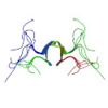
* Ohnishi, S., Güntert, P., Koshiba, S., Tomizawa, T., Akasaka, R., Tochio, T., Sato, M., Inoue, M., Harada, T., Watanabe, S., Tanaka, A., Shirouzu, Kigawa, T. & Yokoyama, S. Solution structure of an atypical WW domain in a novel β-clam-like dimeric form. FEBS Lett. 581, 462-468 (2007).<br>'''PDB [http://www.rcsb.org/pdb/explore.do?structureId=2DWV 2DWV]''' | |* Ohnishi, S., Güntert, P., Koshiba, S., Tomizawa, T., Akasaka, R., Tochio, T., Sato, M., Inoue, M., Harada, T., Watanabe, S., Tanaka, A., Shirouzu, Kigawa, T. & Yokoyama, S. Solution structure of an atypical WW domain in a novel β-clam-like dimeric form. FEBS Lett. 581, 462-468 (2007).<br>'''PDB [http://www.rcsb.org/pdb/explore.do?structureId=2DWV 2DWV]''' | ||
|- | |- | ||
|[[image:2dwv asym r 250.jpg|right|100px|2DWV]] | |[[image:2dwv asym r 250.jpg|right|100px|2DWV]] | ||
* Kuwasako, K., He, F., Inoue, M., Tanaka, A., Sugano, S., Güntert, P., Muto, Y. & Yokoyama, S. Solution structures of the SURP domains and the subunit-assembly mechanism within the splicing factor SF3a complex in 17S U2 snRNP. Structure 14, 1677-1689 (2006).<br>'''PDB [http://www.rcsb.org/pdb/explore.do?structureId=2DT6 2DT6]''' (SURP1)<br>'''PDB [http://www.rcsb.org/pdb/explore.do?structureId=2DT7 2DT7]''' (SURP2-SF3a60) | |* Kuwasako, K., He, F., Inoue, M., Tanaka, A., Sugano, S., Güntert, P., Muto, Y. & Yokoyama, S. Solution structures of the SURP domains and the subunit-assembly mechanism within the splicing factor SF3a complex in 17S U2 snRNP. Structure 14, 1677-1689 (2006).<br>'''PDB [http://www.rcsb.org/pdb/explore.do?structureId=2DT6 2DT6]''' (SURP1)<br>'''PDB [http://www.rcsb.org/pdb/explore.do?structureId=2DT7 2DT7]''' (SURP2-SF3a60) | ||
|- | |- | ||
|} | |} | ||
* ENTH-VHS domain At3g16270(9-135) from ''Arabidopsis thaliana''.<br>López-Méndez, B. & Güntert, P. Automated protein structure determination from NMR spectra. J. Am. Chem. Soc. 128, 13112-13122 (2006).<br>'''PDB [http://www.rcsb.org/pdb/explore.do?structureId=2DCP 2DCP]''' NMR restraints<br>Note: Automated structure determination with FLYA. Structure does not supersede the original deposition 1VDY. | * ENTH-VHS domain At3g16270(9-135) from ''Arabidopsis thaliana''.<br>López-Méndez, B. & Güntert, P. Automated protein structure determination from NMR spectra. J. Am. Chem. Soc. 128, 13112-13122 (2006).<br>'''PDB [http://www.rcsb.org/pdb/explore.do?structureId=2DCP 2DCP]''' NMR restraints<br>Note: Automated structure determination with FLYA. Structure does not supersede the original deposition 1VDY. | ||
Revision as of 10:35, 9 December 2008
NMR protein structures co-authored by P. Güntert.
| * Ohnishi, S., Güntert, P., Koshiba, S., Tomizawa, T., Akasaka, R., Tochio, T., Sato, M., Inoue, M., Harada, T., Watanabe, S., Tanaka, A., Shirouzu, Kigawa, T. & Yokoyama, S. Solution structure of an atypical WW domain in a novel β-clam-like dimeric form. FEBS Lett. 581, 462-468 (2007). PDB 2DWV | |
| * Kuwasako, K., He, F., Inoue, M., Tanaka, A., Sugano, S., Güntert, P., Muto, Y. & Yokoyama, S. Solution structures of the SURP domains and the subunit-assembly mechanism within the splicing factor SF3a complex in 17S U2 snRNP. Structure 14, 1677-1689 (2006). PDB 2DT6 (SURP1) PDB 2DT7 (SURP2-SF3a60) |
- ENTH-VHS domain At3g16270(9-135) from Arabidopsis thaliana.
López-Méndez, B. & Güntert, P. Automated protein structure determination from NMR spectra. J. Am. Chem. Soc. 128, 13112-13122 (2006).
PDB 2DCP NMR restraints
Note: Automated structure determination with FLYA. Structure does not supersede the original deposition 1VDY.
- Rhodanese homology domain At3g16270(175-295) from Arabidopsis thaliana.
López-Méndez, B. & Güntert, P. Automated protein structure determination from NMR spectra. J. Am. Chem. Soc. 128, 13112-13122 (2006). PDB 2DCQ NMR restraints Note: Automated structure determination with FLYA. Structure does not supersede the original deposition 1VEE.
- Src homology domain 2 from the human feline sarcoma oncogene Fes.
López-Méndez, B. & Güntert, P. Automated protein structure determination from NMR spectra. J. Am. Chem. Soc. 128, 13112-13122 (2006). PDB 2DCR NMR restraints Note: Automated structure determination with FLYA. Structure does not supersede the original deposition 1WQU.
- NikA N-terminal fragment.
Yoshida, H., Furuya, N., Lin, Y. J., Güntert, P., Komano, T. & Kainosho, M. PDB 2BA3
- Jurt, S., Aemissegger, A., Güntert, P., Zerbe, O. & Hilvert, D. A photoswitchable miniprotein based on the sequence of avian pancreatic polypeptide. Angew. Chem. Int. Ed. 45, 6297-6300 (2006).
PDB 2H4B NMR restraints (cis-1-PP) 2H3S NMR restraints (cis-1-PP bound to DPC micelles) 2H3T NMR restraints (trans-1-PP bound to DPC micelles)
- Hamada, T., Ito, Y., Abe, T., Hayashi, F., Güntert, P., Inoue, M., Kigawa, T., Terada, T., Shirouzu, M., Yoshida, M., Tanaka, A., Sugano, S., Yokoyama, S. & Hirota, H. Solution structure of the antifreeze-like domain of human sialic acid synthase. Protein Sci. 15, 1010-1016 (2006).
PDB 1WVO NMR restraints
- Aachmann, F. L., Svanem, B. I. G., Güntert, P., Petersen, S. B., Valla, S. & Wimmer, R. NMR structure of the R-module - A parallel β-roll subunit from an Azotobacter vinelandii mannuronan C-5 epimerase. J. Biol. Chem. 281, 7350-7356 (2006).
PDB 2AGM NMR restraints
- Stereo-array isotope labeled (SAIL) maltodextrin-binding protein MBP.
Kainosho, M., Torizawa, T., Iwashita, Y., Terauchi, T., Ono, A. M. & Güntert, P. Optimal isotope labelling for NMR protein structure determinations. Nature 440, 52-57 (2006). PDB 2D21 BMRB 6807
- Stereo-array isotope labeled (SAIL) calmodulin.
Kainosho, M., Torizawa, T., Iwashita, Y., Terauchi, T., Ono, A. M. & Güntert, P. Optimal isotope labelling for NMR protein structure determinations. Nature 440, 52-27 (2006). PDB 1X02 BMRB 6541
- Li, H., Inoue, M., Yabuki, T., Aoki, M., Seki, E., Matsuda, T., Nunokawa, E., Motoda, Y., Kobayashi, A., Terada, T., Shirouzu, M., Koshiba, S., Lin, Y. J., Güntert, P., Suzuki, H., Hayashizaki, Y., Kigawa, T. & Yokoyama, S. Solution structure of the mouse enhancer of rudimentary protein reveals a novel fold. J. Biomol. NMR 32, 329-334 (2005).
PDB 1WWQ
- Scott, A., Pantoja-Uceda, D., Koshiba, S., Inoue, M., Kigawa, T., Terada, T., Shirouzu, M., Tanaka, A., Sugano, S., Yokoyama, S. & Güntert, P. Solution structure of the Src homology 2 domain from the human feline sarcoma oncogene Fes. J. Biomol. NMR 31, 357-361 (2005).
PDB 1WQU BMRB 6331
- Nameki, N., Tochio, N., Koshiba, S., Tochio, N., Inoue, M., Yabuki, T., Aoki, M., Seki, E., Matsuda, T., Saito, M., Ikari, M., Watanabe, M., Terada, T., Shirouzu, M., Yoshida, M., Hirota, H., Tanaka, A., Hayashizaki, Y., Güntert, P., Kigawa, T. & Yokoyama, S. Solution structure of the PWWP domain of the hepatoma-derived growth factor family. Prot. Sci. 14, 756-764 (2005).
PDB 1N27
- Calzolai, L., Lysek, D. A., Pérez, D. R., Güntert, P. & Wüthrich, K. Prion protein NMR structures of chicken, turtle and frog. Proc. Natl. Acad. Sci. USA 102, 651-655 (2005).
PDB 1U3M NMR restraints (chicken PrP(121-225)) 1U5L NMR restraints (turtle PrP(121-225)) 1XU0 NMR restraints (frog PrP(90-222)) BMRB 6269 (chicken PrP(128-242)) 6270 (chicken PrP(25-242)) 6282 (turtle PrP(121-225))
- Lysek, D. A., Schorn, C., Nivon, L. G., Esteve-Moya, V., Christen, B., Calzolai, L., von Schroetter, C., Fiorito, F., Herrmann, T., Güntert, P. & Wüthrich, K. Prion protein NMR structures of cat, dog, pig and sheep. Proc. Natl. Acad. Sci. USA 102, 640-645 (2005).
PDB 1XYJ NMR restraints (cat PrP(121-231)) 1XYK NMR restraints (dog PrP(121-231)) 1XYQ NMR restraints (pig PrP(121-231)) 1XYU NMR restraints (sheep PrP[H168](121-231)) 1Y2S NMR restraints (sheep PrP[R168](121-231)) BMRB 6377 (cat PrP(121-231)) 6378 (dog PrP(121-231)) 6380 (pig PrP(121-231)) 6381 (sheep PrP[H168](121-231)) 6403 (sheep PrP[H168](121-231))
- Pääkkönen, K., Tossavainen, H., Permi, P., Rakkolainen, H., Rauvala, H., Raulo, E., Kilpeläinen, I. & Güntert, P. Solution structures of the first and fourth TSR domains of F-spondin. Proteins 64, 665-672 (2006).
PDB 1SZL (TSR1) 1VEX (TSR4)
- ENTH-VHS domain At3g16270 from Arabidopsis thaliana.
PDB 1VDY BMRB 5928
- Iwai, H., Forrer, P., Plückthun, A. & Güntert, P. NMR solution structure of the monomeric form of the bacteriophage λ capsid stabilizing protein gpD. J. Biomol. NMR 31, 351-356 (2005).
PDB 1VD0 BMRB 4936
- Pantoja-Uceda, D., López-Méndez, B., Koshiba, S., Inoue, M., Kigawa, T., Terada, T., Shirouzu, M., Tanaka, A., Seki, M., Shinozaki, K., Yokoyama, S. & Güntert, P. Solution structure of the rhodanese homology domain At4g01050(175-295) from Arabidopsis thaliana. Prot. Sci. 14, 224-230 (2005).
PDB 1VEE BMRB 5929
- Nameki, N., Yoneyama, M., Koshiba, S., Tochio, N., Inoue, M., Seki, E., Matsuda, T., Tomo, Y., Harada, T., Saito, K., Kobayashi, N., Yabuki, T., Aoki, M., Nunokawa, E., Matsuda, N., Sakagami, N., Terada, T., Shirouzu, M., Yoshida, M., Hirota, H., Osanai, T., Tanaka, A., Arakawa, T., Carninci, P., Kawai, J., Hayashizaki, Y., Kinoshita, K., Güntert, P., Kigawa, T. & Yokoyama, S. Solution structure of the RWD domain of the mouse GCN2 protein. Prot. Sci. 13, 2089-2100 (2004).
PDB 1UKX
- Fernández, C., Hilty, C., Wider, G., Güntert, P. & Wüthrich, K. NMR Structure of the integral membrane protein OmpX. J. Mol. Biol. 336, 1211-1221 (2004).
PDB 1Q9G BMRB 4936
- FHA domain of mouse afadin 6.
PDB 1WLN
- Vanwetswinkel, S., Kriek, J., Andersen, G. R., Güntert, P., Dijk, J., Canters, G. W. & Siegal, G. Solution structure of the 162 residue C-terminal domain of the human elongation factor 1Bγ. J. Biol. Chem. 278, 43443-43451 (2003).
PDB 1PBU NMR restraints BMRB 5628
- Hilge, M., Siegal, G., Vuister, G. W., Güntert, P., Gloor, S. M. & Abrahams, J. P. ATP-induced conformational changes of the nucleotide-binding domain of Na,K-ATPase. Nat. Struct. Biol. 10, 468-474 (2003).
PDB 1MO7 (free form) 1MO8 (ATP-bound form) BMRB 5577 (free form) 5576 (ATP-bound form)
- Lührs, T., Riek, R., Güntert, P. & Wüthrich, K. NMR structure of the human doppel protein. J. Mol. Biol. 326, 1549-1557 (2003).
PDB 1LG4 NMR restraints BMRB 5145
- Zahn, R., Güntert, P., von Schroetter, C. & Wüthrich, K. NMR structure of a human prion protein with two disulfide bridges. J. Mol. Biol. 326, 225-234 (2003).
PDB 1H0L NMR restraints BMRB 5378
- Enggist, E., Thöny-Meyer, L., Güntert, P. & Pervushin, K. NMR structure of the heme chaperone CcmE reveals a novel functional motif. Structure 10, 1551-1557 (2002).
PDB 1SR3 NMR restraints
- Lee, D., Damberger, F. D., Peng, G., Horst, R., Güntert, P., Nikonova, L., Leal, W. S. & Wüthrich, K. NMR structure of the unliganded Bombyx mori pheromone-binding protein at physiological pH. FEBS Lett. 531, 314-318 (2002).
PDB 1LS8
- Ellgaard, L., Bettendorff, P., Braun, D., Herrmann, T., Fiorito, F., Jelesarov, I., Herrmann, T., Güntert, P., Helenius, A. & Wüthrich, K. NMR structures of 36 and 73-residue fragments of the calreticulin P-domain. J. Mol. Biol. 322, 773-784 (2002).
PDB 1K9C NMR restraints (residues 189-261) 1K91 (residues 221-256) BMRB 5204 (residues 189-261) 5205 (residues 221-256)
- Miura, T., Klaus, W., Ross, A., Güntert, P. & Senn, H. The NMR structure of the class I human ubiquitin-conjugating enzyme 2b. J. Biomol. NMR 22, 89-92 (2002).
PDB 1JAS NMR restraints BMRB 5038
- Horst, R., Damberger, F., Luginbühl, P., Güntert, P., Peng, G., Nikonova, L., Leal, W. S. & Wüthrich, K. NMR structure reveals intramolecular regulation mechanism for pheromone binding and release. Proc. Natl. Acad. Sci. USA 98, 14374-14379 (2001).
PDB 1GM0 NMR restraints BMRB 4849
- Ellgaard, L., Riek, R., Herrmann, T., Güntert, P., Braun, D., Helenius, A. & Wüthrich, K. NMR structure of the calreticulin P-domain. Proc. Natl. Acad. Sci. USA 98, 3133-3138 (2001).
PDB 1HHN BMRB 4878
- Calzolai, L., Lysek, D. A., Güntert, P., von Schroetter, C., Riek, R., Zahn, R. & Wüthrich, K. NMR structures of three single-residue variants of the human prion protein. Proc. Natl. Acad. Sci. USA 97, 8340-8345 (2000).
PDB 1E1G NMR restraints (variant M166V) 1E1P NMR restraints (variant S170N) 1E1U NMR restraints (variant R220K) BMRB 4736 (variant M166V) 4620 (variant R220K)
- Riek, R., Prêcheur, P., Wang, Y., Mackay, E. A., Wider, G., Güntert, P., Liu, A., Kägi, J. H. R. & Wüthrich, K. NMR structure of the sea urchin (Strongylocentrotus purpuratus) metallothionein MTA. J. Mol. Biol. 291, 417-428 (1999).
PDB 1QJK NMR restraints (alpha domain) 1QJL NMR restraints (beta domain) BMRB 4363
- Pellecchia, M., Güntert, P., Glockshuber, R. & Wüthrich, K. The NMR solution structure of the periplasmic chaperone FimC. Nature Struct. Biol. 5, 885-890 (1998).
PDB 1BF8 BMRB 4070
- Ottiger, M., Zerbe, O., Güntert, P. & Wüthrich, K. The NMR solution conformation of unligated human cyclophilin A. J. Mol. Biol. 272, 64-81 (1997).
PDB 1OCA NMR restraints
- Antuch, W., Güntert, P. & Wüthrich, K. Ancestral beta gamma-crystallin precursor structure in a yeast killer toxin, Nature Struct. Biol. 3, 662-665 (1996).
PDB 1WKT NMR restraints BMRB 5255
- Antuch, W., Güntert, P., Billeter, M., Hawthorne, T., Grossenbacher, H. & Wüthrich, K. NMR solution structure of the recombinant tick anticoagulant protein (rTAP), a factor Xa inhibitor from the tick Ornithodoros moubata. FEBS Lett. 352, 251-257 (1994).
PDB 1TAP
- Berndt, K. D., Güntert, P. & Wüthrich, K. The NMR solution structure of dendrotoxin K from the venom of Dendroaspis polylepis polylepis. J. Mol. Biol. 234, 735-750 (1993).
PDB 1DTK NMR restraints
- Szyperski, T., Güntert, P., Stone, S. R. & Wüthrich, K. The NMR solution structure of hirudin(1-51) and comparison with corresponding three-dimensional structures determined using the complete 65-residue hirudin polypeptide chain. J. Mol. Biol. 228, 1193-1205 (1992).
PDB 1HIC NMR restraints
- Berndt, K. D., Güntert, P., Orbons, L. P. M. & Wüthrich, K. Determination of a high-quality NMR solution structure of the bovine pancreatic trypsin inhibitor (BPTI) and comparison with three crystal structures. J. Mol. Biol. 227, 757-775 (1992).
PDB 1PIT NMR restraints
- Güntert, P., Qian, Y. Q., Otting, G., Müller, M., Gehring, W. J. & Wüthrich K. Structure determination of the Antp(C39->S) homeodomain from nuclear magnetic resonance data in solution using a novel strategy for the structure calculation with the programs DIANA, CALIBA, HABAS and GLOMSA. J. Mol. Biol. 217, 531-540 (1991).
PDB 2HOA
